

Improving Detection: AI-Assisted Cervical Imaging
- May 19
- 4 min read

From Image to Impact
AI-assisted cervical imaging can support better care. A clearer image can lead to an earlier diagnosis, and more timely treatment.
And in many cases, that can mean the difference between a condition that can be treated and one that is much harder to manage.
Improving detection starts with the image
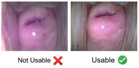
Artificial intelligence (AI) can help identify precancerous changes in the cervix, but its performance often depends on the quality of a single image.
In many clinics around the world, a cervical cancer diagnosis can depend on a single image.
If that image is blurry or poorly lit, early signs of disease can be missed. In settings where patients may not return for follow-up care, a missed diagnosis can have lasting consequences.
Last month, we explored how the Callascope is helping remove barriers to screening by making the experience more accessible and patient centered. But expanding access is only the first step. Once a patient is screened, the next challenge is ensuring that what clinicians see, and what algorithms analyze, is accurate and reliable.
At the Duke Center for Global Women’s Health Technologies (GWHT), researchers are working to address this next step by rethinking how images are captured, interpreted, and used to support early detection.
A Different Starting Point
The work at GWHT begins one step before the image is analyzed by an algorithm.
“Before we even get to diagnosis, we have to ask whether the images themselves are usable,” says Lillian Ekem, a GWHT PhD candidate whose research focuses on image quality.
In practice, many cervical images suffer from blur, glare, or incomplete views of the cervix. These issues can make it difficult for clinicians to interpret what they see and can result in algorithms learning from incorrect or misleading information.
“If something looks abnormal, we need to know whether that’s actually disease or just the way the image was captured,” Ekem explains.
Prior studies from the team showed that image blur reduced provider confidence in interpreting cervical images. Blur and shadow also affected how consistently AI algorithms made predictions, highlighting the importance of image quality for both clinical interpretation and AI-assisted diagnosis.
To address this, the team developed AI tools that quickly assess whether an image is interpretable so providers can determine whether an image should be retaken or whether the imaging device should be repositioned.
Designing with Intention
Glare, shadows, and natural anatomical variation can affect how an algorithm interprets an image. To reduce that risk, the team is developing ways to clean and standardize images, so clinicians and algorithms can focus on what matters.
For Ekem, this work reflects a broader shift in how the team approaches technology.
“Technology alone can’t fix inequality,” she says. “We have to be intentional about how we build and use it.” Instead of focusing only on diagnostic algorithms, the group is looking at the system, with consideration for how images are captured, processed, and used by providers.
For example, in many low-resource areas, cervical image capture may be administered by a general practitioner without the specialized knowledge to confidently interpret an image alone. For non-specialists, the instant algorithmic feedback Ekem is developing can serve as a training tool, offering guidance, increased skill, and confidence in capturing and interpreting quality images.
Jessica De Souza, a GWHT Postdoctoral Fellow, also works in developing the AI tools within the whole context of the clinical workflow and its complex variations.
“There are features in the image that can influence predictions that have nothing to do with disease, like blur, shadows, and natural anatomical variation” she explains . “We want the algorithm to focus on biologically meaningful features rather than artifacts introduced during image capture.”
The team is developing AI tools that identify clinically important regions of the cervix while accounting for differences in anatomy, image quality and imaging devices.
The goal is to make AI-assisted cervical screening more accurate, consistent, and better assist in real-world clinical settings. “We want the provider to make the decision,” De Souza says. “The AI is there to guide, not decide.”
Seeing More Than a Snapshot
Beyond image quality and anatomical variability, another challenge is how cervical images are acquired over time.
Traditional screening methods rely on applying contrast agents to the cervix and observing how the tissue looks over time. Areas that change in appearance may indicate abnormal cells. This approach provides a more complete picture: “When you can see how the tissue evolves over time, you have much more information,” Ekem explains.
Expanding Access, Thoughtfully
A key part of this work is improving access.
Low-cost imaging devices, like the GWHT-developed, speculum-free Callascope, are making it possible to bring cervical imaging into settings where traditional equipment is not available (see last month’s article for more). Images from with these devices can be analyzed using a smartphone app.
These tools are part of a shift toward care models that meet patients where they are.
As these devices become more widely used, ensuring the quality of the images they produce becomes even more important. The GWHT Cervix Team’s work helps make sure these images are usable for both clinical decisions and for future research.


Comments